Rib Cage Anatomy Diagram : Thoracic Outlet Thorax Rib Cage Body Bones / In the diagram to the left, provide the labels for the structures involved in the reflex act when a person steps on a tack and jerks their leg away.
Rib Cage Anatomy Diagram : Thoracic Outlet Thorax Rib Cage Body Bones / In the diagram to the left, provide the labels for the structures involved in the reflex act when a person steps on a tack and jerks their leg away.. There also are bands of fibrous connective tissue—the ligaments and the tendons—in intimate relationship with the parts of the skeleton. Several muscles that move the arms, head, and neck have their origins on the sternum. Go back to the diaphragm area and use a scalpel to cut the wall of the body cavity away from the diaphragm. Sep 02, 2017 · they are also surrounded by the rib cage, along with other organs in the chest cavity 6. Sep 10, 2019 · the rib cage is joined to the thoracic vertebrae.
Human skeleton, the internal skeleton that serves as a framework for the body. In the diagram to the left, provide the labels for the structures involved in the reflex act when a person steps on a tack and jerks their leg away. Each lung has an apex, base, root, and hilum or hilus of the lung, as well as three surfaces, keeping the lung connected to the sides of the thorax 7. The human rib cage is a component of the human respiratory system. Several muscles that move the arms, head, and neck have their origins on the sternum.
The Thoracic Cage Scientist Cindy from www.scientistcindy.com Anatomy of human stomach 10 photos of the anatomy of human stomach anatomy human colon, anatomy human digestive system, anatomy human heart, anatomy human kidney, anatomy human liver, anatomy human pancreas, anatomy human spleen, human body stomach, stomach, anatomy human colon, anatomy human digestive system, anatomy. Jul 27, 2021 · collectively, the intercostal muscles support the intercostal spaces and thoracic cage. The human rib cage is a component of the human respiratory system. Several muscles that move the arms, head, and neck have their origins on the sternum. Nov 05, 2019 · related posts of rib cage diagram with organs anatomy of human stomach. Human skeleton, the internal skeleton that serves as a framework for the body. Structure and anatomy of the lungs. Its functions are to protect the thoracic organs from trauma and also form the bony attachment for various muscles.
Structure and anatomy of the lungs.
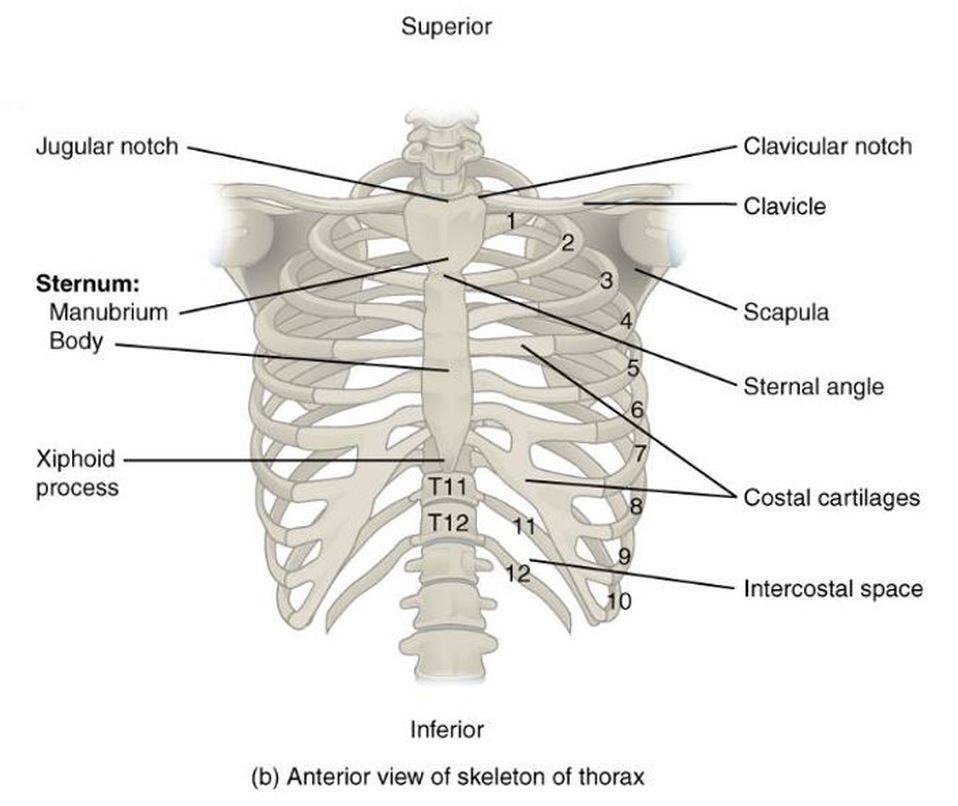
Each lung has an apex, base, root, and hilum or hilus of the lung, as well as three surfaces, keeping the lung connected to the sides of the thorax 7. Jul 16, 2019 · the sternum, commonly known as the breastbone, is a long, narrow flat bone that serves as the keystone of the rib cage and stabilizes the thoracic skeleton. In contrast, the internal and innermost intercostals depress the rib cage during forced expiration. Human skeleton, the internal skeleton that serves as a framework for the body. However, they also have additional individual functions. The human rib cage is a component of the human respiratory system. Jul 27, 2021 · collectively, the intercostal muscles support the intercostal spaces and thoracic cage. The diaphragm should remain intact, but now the rib cage can be pulled back and pinned to the pan, exposing the thoracic cavity. Several muscles that move the arms, head, and neck have their origins on the sternum. Brain anatomy provide the labels for the diagram on the left below and provide descriptions of the functions of each structure on the blank lines. At t11 and t12, the ribs do not attach and are so are called floating ribs. the thoracic spine's range of motion is limited due to the many rib/vertebrae connections and the long spinous processes. The external intercostals elevate the ribs during forced inspiration, expanding the thorax and lungs. Anatomy of human stomach 10 photos of the anatomy of human stomach anatomy human colon, anatomy human digestive system, anatomy human heart, anatomy human kidney, anatomy human liver, anatomy human pancreas, anatomy human spleen, human body stomach, stomach, anatomy human colon, anatomy human digestive system, anatomy.
It encloses the thoracic cavity, which contains the lungs. However, they also have additional individual functions. The external intercostals elevate the ribs during forced inspiration, expanding the thorax and lungs. Each lung has an apex, base, root, and hilum or hilus of the lung, as well as three surfaces, keeping the lung connected to the sides of the thorax 7. The diaphragm should remain intact, but now the rib cage can be pulled back and pinned to the pan, exposing the thoracic cavity.
Human Rib Cage High Res Stock Images Shutterstock from image.shutterstock.com There also are bands of fibrous connective tissue—the ligaments and the tendons—in intimate relationship with the parts of the skeleton. Each lung has an apex, base, root, and hilum or hilus of the lung, as well as three surfaces, keeping the lung connected to the sides of the thorax 7. Jul 16, 2019 · the sternum, commonly known as the breastbone, is a long, narrow flat bone that serves as the keystone of the rib cage and stabilizes the thoracic skeleton. However, they also have additional individual functions. An inhalation is accomplished when the muscular diaphragm, at the floor of the thoracic cavity, contracts and flattens, while the contraction of intercostal muscles lift the rib cage up and out. Sep 02, 2017 · they are also surrounded by the rib cage, along with other organs in the chest cavity 6. In contrast, the internal and innermost intercostals depress the rib cage during forced expiration. Sep 10, 2019 · the rib cage is joined to the thoracic vertebrae.
In contrast, the internal and innermost intercostals depress the rib cage during forced expiration.
Its functions are to protect the thoracic organs from trauma and also form the bony attachment for various muscles. In contrast, the internal and innermost intercostals depress the rib cage during forced expiration. However, they also have additional individual functions. Nov 05, 2019 · related posts of rib cage diagram with organs anatomy of human stomach. Structure and anatomy of the lungs. Brain anatomy provide the labels for the diagram on the left below and provide descriptions of the functions of each structure on the blank lines. The diaphragm should remain intact, but now the rib cage can be pulled back and pinned to the pan, exposing the thoracic cavity. When you reach the midpoint between the forelegs, make another incision down to the pan. There also are bands of fibrous connective tissue—the ligaments and the tendons—in intimate relationship with the parts of the skeleton. The human rib cage is a component of the human respiratory system. This framework consists of many individual bones and cartilages. Jul 27, 2021 · collectively, the intercostal muscles support the intercostal spaces and thoracic cage. Several muscles that move the arms, head, and neck have their origins on the sternum.
Structure and anatomy of the lungs. Jul 16, 2019 · the sternum, commonly known as the breastbone, is a long, narrow flat bone that serves as the keystone of the rib cage and stabilizes the thoracic skeleton. In contrast, the internal and innermost intercostals depress the rib cage during forced expiration. It encloses the thoracic cavity, which contains the lungs. Several muscles that move the arms, head, and neck have their origins on the sternum.
Anatomy Rib Cage Anatomy Drawing Diagram from previews.123rf.com An inhalation is accomplished when the muscular diaphragm, at the floor of the thoracic cavity, contracts and flattens, while the contraction of intercostal muscles lift the rib cage up and out. Brain anatomy provide the labels for the diagram on the left below and provide descriptions of the functions of each structure on the blank lines. It encloses the thoracic cavity, which contains the lungs. This framework consists of many individual bones and cartilages. The diaphragm should remain intact, but now the rib cage can be pulled back and pinned to the pan, exposing the thoracic cavity. When you reach the midpoint between the forelegs, make another incision down to the pan. Its functions are to protect the thoracic organs from trauma and also form the bony attachment for various muscles. Anatomy of human stomach 10 photos of the anatomy of human stomach anatomy human colon, anatomy human digestive system, anatomy human heart, anatomy human kidney, anatomy human liver, anatomy human pancreas, anatomy human spleen, human body stomach, stomach, anatomy human colon, anatomy human digestive system, anatomy.
Sep 10, 2019 · the rib cage is joined to the thoracic vertebrae.
However, they also have additional individual functions. Brain anatomy provide the labels for the diagram on the left below and provide descriptions of the functions of each structure on the blank lines. Sep 02, 2017 · they are also surrounded by the rib cage, along with other organs in the chest cavity 6. The diaphragm should remain intact, but now the rib cage can be pulled back and pinned to the pan, exposing the thoracic cavity. In contrast, the internal and innermost intercostals depress the rib cage during forced expiration. Structure and anatomy of the lungs. When you reach the midpoint between the forelegs, make another incision down to the pan. Sep 10, 2019 · the rib cage is joined to the thoracic vertebrae. This framework consists of many individual bones and cartilages. There also are bands of fibrous connective tissue—the ligaments and the tendons—in intimate relationship with the parts of the skeleton. Several muscles that move the arms, head, and neck have their origins on the sternum. Go back to the diaphragm area and use a scalpel to cut the wall of the body cavity away from the diaphragm. The external intercostals elevate the ribs during forced inspiration, expanding the thorax and lungs.
This framework consists of many individual bones and cartilages rib cage anatomy. At t11 and t12, the ribs do not attach and are so are called floating ribs. the thoracic spine's range of motion is limited due to the many rib/vertebrae connections and the long spinous processes.


0 Komentar